The University of Southern Queensland (USQ) is committed to advancing the use …
The University of Southern Queensland (USQ) is committed to advancing the use of open textbooks in higher education. This textbook is a tool to support first year anatomy and physiology courses taught in Australia, aiming to provide students with an increased access to free, high-quality learning materials.
The material in this textbook is largely based on OpenStax’s Anatomy & Physiology textbook, however, has been modified for Australian course curriculum.
Table of Contents I. Levels of Organisation, Homeostasis and Nomenclature II. Cells and Reproduction III. Tissues, Organs, Systems IV. Integumentary System V. Blood VI. Cardiovascular System VII. Lymphatic System and Immunity VIII. Respiratory System IX. Muscle System X. Skeletal System XI. Musculoskeletal System XII. Digestive System XIII. Nervous System XIV. Endocrine System XV. Reproductive System XVI. Pregnancy and Human Development XVII. Urinary System
The University of Southern Queensland (USQ) is committed to advancing the use …
The University of Southern Queensland (USQ) is committed to advancing the use of open textbooks in higher education. This textbook is a tool to support first year anatomy and physiology courses taught in Australia, aiming to provide students with an increased access to free, high-quality learning materials.
The gall bladder stores bile produced in the liver. Bile is important …
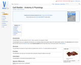
The gall bladder stores bile produced in the liver. Bile is important in the digestion of lipids. The gall bladder forms as an outgrowth of the bile duct, as a secondary hollow at the posterior edge of the original hepatic rudiment. The cystic duct joins the common bile duct which enters the duodenum at the major duodenal papillae (with the pancreatic duct) on the dorsal surface of the duodenum.
The air in the alveoli is renewed regularly, thanks to the ventilation …
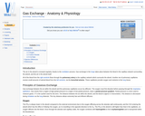
The air in the alveoli is renewed regularly, thanks to the ventilation process. Gas exchange in the lungs takes place between the blood in the capillary network surrounding the alveoli, and the air in the alveoli itself.
Presents the anatomy, physiology, biochemistry, biophysics, and bioengineering of the gastrointestinal tract …
Presents the anatomy, physiology, biochemistry, biophysics, and bioengineering of the gastrointestinal tract and associated pancreatic, liver, and biliary systems. Emphasis on the molecular and pathophysiological basis of disease where known. Covers gross and microscopic pathology and clinical aspects. Formal lectures given by core faculty, with some guest lectures by local experts. Selected seminars conducted by students with supervision of faculty. Permission of instructor required. (Only HST students may register under HST.120, graded P/D/F.) The most recent knowledge of the anatomy, physiology, biochemistry, biophysics, and bioengineering of the gastrointestinal tract and the associated pancreatic, liver and biliary tract systems is presented and discussed. Gross and microscopic pathology and the clinical aspects of important gastroenterological diseases are then presented, with emphasis on integrating the molecular, cellular and pathophysiological aspects of the disease processes to their related symptoms and signs.
Gastrulation is the process of forming the three germ layers; ectoderm, mesoderm …
Gastrulation is the process of forming the three germ layers; ectoderm, mesoderm and endoderm. It is achieved through a series of highly coordinated cell movements. Cells that will form the endodermal and mesodermal organs are brought inside the embryo, whilst cells that will form ectoderm move to spread out over the outside of the embryo.
An integrated course stressing the principles of biology. Life processes are examined …
An integrated course stressing the principles of biology. Life processes are examined primarily at the organismal and population levels. Intended for students majoring in biology or for non-majors who wish to take advanced biology courses.
The endoderm will form the lining of the gut and the organs …
The endoderm will form the lining of the gut and the organs that develop from it. Splanchnic mesoderm surrounds the endoderm and orginates from the lateral plate mesoderm. It will form the smooth muscle of the gut that are used in peristalsis.
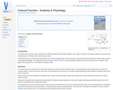
The Guttural Pouch is present only in members of the order Perissodactyla …
The Guttural Pouch is present only in members of the order Perissodactyla (nonruminant ungulates: horses, tapirs, rhinoceros) and another small band of small mammals including Hyraxes, certain bats and a South American mouse.
Hair germs begin from an aggregation of keratinocytes in the stratum basale …
Hair germs begin from an aggregation of keratinocytes in the stratum basale of the epidermis. The initiating factor is the underlying dermal fibroblast cells. The keratinocytes elongate, divide and relocate to the dermis. Dermal fibroblasts then form a dermal papilla beneath the hair germ. This causes stimulation of the basal stem cells to up-regulate their cycle, producing cells that will keratinise and form the hair shaft. Two swellings form on the shaft, one containing stem cells for follicle regeneration, the other becomes a sebaceous gland which will secrete sebum onto the hair shaft. The follicles develop from an ectodermal bud which invades the mesenchyme during embryonic development. The mesoderm also condenses during the development creating an outer mesodermal component to the embedded part of the hair.
The formation of the mammalian heart is a fairly complex process. It …
The formation of the mammalian heart is a fairly complex process. It begins when angiogenic mesodermal cells in the cardiogenic plate coalesce to form the endocardial tubes. The endocardial tubes then fuse to form a single duct, the cardiac tube. This undergoes a process of distension, folding and septation and a four chambered, dual circuit pump is formed . The simple heart seen in fish or amphibians forms via the same path but development ceases at an earlier stage.
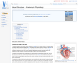
The heart is located in the thoracic cavity in between the lungs, …
The heart is located in the thoracic cavity in between the lungs, 60% of it lying to the left of the median plane. The hearts lateral projection extends from rib 3 to 6. Most of the hearts surface is covered by the lungs and in juveniles it is bordered cranially by the thymus. Caudally the heart extends as far as the diaphragm. Variations in position and size exist among individuals depending on species, breed, age, fitness and pathology. Roughly speaking, the heart is responsible for about 0.75% of the bodyweight.
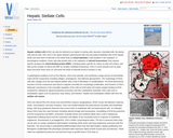
Hepatic stellate cells (HSC) can also be referred to as vitamin A-storing …
Hepatic stellate cells (HSC) can also be referred to as vitamin A-storing cells, lipocytes, interstitial cells, fat-storing cells and Ito cells. HSC exist in the space between parenchymal cells and sinusoidal endothelial cells of the hepatic lobule and store 80% of retinoids in the whole body as retinyl palmitate in lipid droplets in the cytoplasm. In physiological conditions, these cells play pivotal roles in the regulation of retinoid homeostasis; they express specific receptors for retinol-binding protein (RBP), a binding protein specific for retinol, on their cell surface, and take up the complex of retinol and RBP by receptor-mediated endocytosis.
The hind brain is also called the rhombencephalon and is the brain …
The hind brain is also called the rhombencephalon and is the brain stem that provides the connection between the spinal cord and the rest of the brain. The hind brain contains many vital structures including the Medulla Oblongata, the Pons (the link between the cerebellum, forebrain and mid-brain) and the majority of the cranial nerves, III to XII. In general the brain stem governs essential functions that are carried out sub-consciously via reflexes.
Hindgut fermenters are evolved to eat a herbivorous diet. Such a diet …
Hindgut fermenters are evolved to eat a herbivorous diet. Such a diet includes large quantities of insoluble plant carbohydrates, such as cellulose. Mammals cannot digest these insoluble carbohydrates as they lack the essential enzymes, such as cellulase. However it is important that they do digest these carbohydrates as there is insufficient quantity of soluble carbohydrates in plant material. Some microbes do have the enzymes to digest these insoluble carbohydrates and so hindgut fermenters hold a symbiotic relationship with these microbes. Hindgut fermenters have anatomical adaptations to allow for an expanded microbial population. The products of fermentation are volatile fatty acids. It is important to supply a source of fibre in their diet as it stimulates peristalsis in the gut and prevents a build up of gas.
The hoof is defined from a physiologic perspective as the modified skin …
The hoof is defined from a physiologic perspective as the modified skin covering the tip of the digit and all enclosed structures. The hoof provides protection to the distal limb and is formed by keratinisation of the epithelial layer and modification of the underlying dermis. The keratin in the epidermis, when thickened and cornified, is referred to as horn. Horn makes up the outer surface if the hoof and is particularly resistant to mechanical and chemical damage.
The keratin in the epidermis, when cornified and thickened, is referred to …
The keratin in the epidermis, when cornified and thickened, is referred to as horn. Horn is particulary resistant to mechanical and chemical damage. The dermis of horn gives the structures their 3-D structure and shape. Cattle, some sheep, goats and antelope posess horns and these are permanent organs. Breeds without horns are termed polled breeds. Deer posess antlers, which are temporary organs that develop during the rutting season and are then shed.
This course will provide the student with an overview of the body …
This course will provide the student with an overview of the body from a systemic perspective. Each unit will focus on one system, or network of organs that work together to perform a particular function. At the end of this course, the student will review the ways in which the systems overlap, as well as discuss current body imaging techniques and learn how to correctly interpret the images in order to put our newly-gained anatomical knowledge to practical use. Upon successful completion of this course, the student will be able to: identify gross and microscopic anatomy and explain interactions of the major organ systems in the human body; perform and analyze experiments in human anatomy (virtual); use language necessary to appropriately describe human anatomy; explain and identify how structure and function complement each other; describe how anatomy relates to medical situations in healthy and diseased states. (Biology 302)
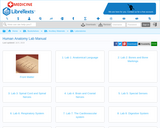
5: Lab 5: Special Senses 5.1: Lab 5: Special Senses 5.2: Pre-lab 5 5.3: Lab Activities 5.4: Post-Lab 5 Questions
6: Lab 6: Respiratory System 6.1: Lab 6: Respiratory System 6.2: Lab Activities 6.3: Post-Lab 6 Questions
7: Lab 7: The Cardiovascular system 7.1: Lab 7: The Cardiovascular system 7.2: Pre-lab 7 7.3: Lab Activities 7.4: Post-Lab 7 Questions
8: Lab 8: Digestive System 8.1: Lab 8: Digestive System 8.2: Pre-Lab 8 8.3: Lab Activities 8.4: Post-Lab 8 Questions
9: Lab 9: Urinary and Reproductive Systems 9.1: Lab 9: Urinary and Reproductive Systems 9.2: Pre-lab 9 9.3: Lab Activities 9.4: Post-Lab 9 Questions
10: Lab 10: The Muscular and Integumentary systems 10.1: Lab 10: The Muscular and Integumentary systems 10.2: Pre-Lab 10 10.3: Lab Activities 10.4: Post-Lab 10 Questions
No restrictions on your remixing, redistributing, or making derivative works. Give credit to the author, as required.
Your remixing, redistributing, or making derivatives works comes with some restrictions, including how it is shared.
Your redistributing comes with some restrictions. Do not remix or make derivative works.
Most restrictive license type. Prohibits most uses, sharing, and any changes.
Copyrighted materials, available under Fair Use and the TEACH Act for US-based educators, or other custom arrangements. Go to the resource provider to see their individual restrictions.