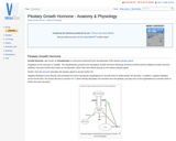
Osmosis is the passive movement of water across a semi permeable membrane. …
Osmosis is the passive movement of water across a semi permeable membrane. It occurs in the opposite direction to diffusion of ions. Water moves from a region of low solute concentration and therefore high water concentration to a region of high solute concentration and low water concentration.
The ovary is the female Gonad homologous to the male Testes. It …
The ovary is the female Gonad homologous to the male Testes. It is usually a paired organ in domestic species, but in the bird only the left Ovary is present. The structures found within the ovary are undergoing constant changes throughout the oestrus cycle from the Follicles containing Oocytes, to the formation of Corpus Haemorrhagicum, Corpus Luteum, and finally Corpus Albicans. Ovaries are ellipsoidal in shape with an irregular surface due to the projection of dominant follicles and corpora lutea. These irregularities are absent in the mare due to the cortex and medulla being reversed with ovulation only occuring from the ovulation fossa. They are greatest in Polytocious animals such as the sow due to many dominant follicles, and so corpora lutea, developing at once.
The Oviduct is the tube that links the ovary to the uterus …
The Oviduct is the tube that links the ovary to the uterus and which the ovulated oocyte travels down to become fertilised by sperm present in the female tract. It is also refered to as the Fallopian tube, Uterine tube or Ovarian tube.
The Peripheral Nervous System is made up of cranial and spinal nerves. …
The Peripheral Nervous System is made up of cranial and spinal nerves. Spinal nerves are named after the vertebra immediately above it, except for cervical vertebra. There are 7 cervical vertebrae and 8 cervical spinal nerves. The peripheral nervous system can be divided into the somatic nervous system and autonomic nervous system.
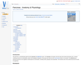
The pancreas is a tubuloalveolar gland and has exocrine and endocrine tissues. …
The pancreas is a tubuloalveolar gland and has exocrine and endocrine tissues. The exocrine is the larger of the two parts and secretes pancreatic juice; a solution containing enzymes for carbohydrate, protein and triacylglycerol digestion. Pancreatic juice drains into the small intestine where it is functional. The endocrine part secretes hormones for the regulation of blood glucose concentration, including insulin, glucagon and somatostatin. The functional units of the endocrine part are the islets of Langerhans.
The paranasal sinuses are ventilated spaces connected to the nasal cavity. They …
The paranasal sinuses are ventilated spaces connected to the nasal cavity. They develop as blind ending pouches between the lamina of the bones of the skull.
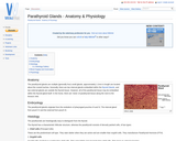
The parathyroid glands are multiple (generally four) small glands, approximately 1-2mm in …
The parathyroid glands are multiple (generally four) small glands, approximately 1-2mm in length are located about the cranial trachea. Generally, there are two internal glands embedded within the thyroid Glands, and two external glands are outside the thyroid tissue. However, all of the parathyroid tissue may be embedded within the thyroid gland itself. In the horse, there are 'nests' of parathyroid tissue along the neck to the thoracic inlet.
Nerves of the peripheral nervous system (PNS) are composed of numerous bundles …
Nerves of the peripheral nervous system (PNS) are composed of numerous bundles of nerve fibers that are surrounded by connective tissue. This connective tissue also contains a specific layer that is specialised to neurons; the peri-neurium. The outer layer of this connective tissue is called the epineurium and it surrounds both the perineurium and the nerve itself. Individual neurons found within each bundle are surrounded by the endoneurium.
The peritoneum is the serous membrane that lines the abdominal cavity. It …
The peritoneum is the serous membrane that lines the abdominal cavity. It lies directly beneath the abdominal musculature (rectus abdominis and transverse abdominis). It is a type of loose connective tissue and is covered by mesothelium. Extensions of the peritoneum form the mesenteries, omenta and ligaments that support the abdominal contents. The peritoneum produces fluid to lubricate abdominal viscera. The peritoneum also enhances immune responses and walls off infection in the abdomen to prevent peritonitis.
The pharynx is part of both the respiratory and digestive system. Both …
The pharynx is part of both the respiratory and digestive system. Both systems have entrances to the pharynx but they are separated from each other by the soft palate. During exercise or during respiratory distress, the mouth can be used as an additional opening of the respiratory system and then the oropharynx also becomes an air-way.
The phospholipid bilayer is the fundamental structure which makes up the cell …
The phospholipid bilayer is the fundamental structure which makes up the cell membrane. It is made of 2 sheets of phospholipid molecules which are said to have hydrophillic heads and hydrophobic tails. Therefore molecules on opposite sheets face back to back to protect their hydrophobic area from the surrounding intra or extracellular fluid. This creates a region inside the membrane which is hydrophobic.
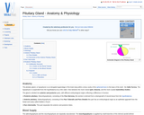
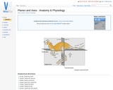
The pituitary gland, or hypophysis is an elongated appendage of the brain …
The pituitary gland, or hypophysis is an elongated appendage of the brain lying within a bony cavity of the sphenoid bone in the base of the skull - the Sella Turcica. The hypophysis is suspended from the hypothalamus by a thin stalk. It lies between the more rostral optic chiasma, and the more caudal mammillary bodies.
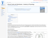
The surface of the inner wall of all of the body cavities …
The surface of the inner wall of all of the body cavities is lined by a serous membrane which consists of a single layer of flat epithelium with a thin underlying propria (connective tissue). Within the thoracic cavity, this is known as the pleura. The visceral pleura which coats the outer surface of the lung is derived from the splanchnic mesoderm. The parietal pleura lining the thoracic cavity is derived from somatic mesoderm. The pleural cavity is a potential space between the two areas of pleural membrane, which normally are adhesed to each other.
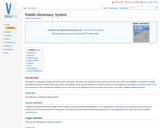
The rabbit is a monogastric hindgut fermenter and is a herbivore. Microbes …
The rabbit is a monogastric hindgut fermenter and is a herbivore. Microbes in the hindgut produce volatile fatty acids (VFAs) which are available to the animal for energy. Microbes also produce vitamins and protein, which are available only in minimal quantities as they are produced in the hindgut (see advantages and disadvantages of hid gut fermentation). Most microbial fermentation occurs in the caecum (as opposed to the horse where most occurs in the colon). Rabbits usually eat at dusk.
The rectum lies between the terminal portion of the descending colon and …
The rectum lies between the terminal portion of the descending colon and anus. It is empty most of the time, except after the mass movements of the large intestine which move faeces into the rectum. This stimulates defeaction, which may happen when an animal is frightened.
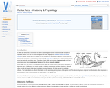
A reflex arc represents a mechanism by which a physiological function is …
A reflex arc represents a mechanism by which a physiological function is automatically managed or regulated. Reflex arcs can be found throughout the body, ranging from skeletal muscles to smooth muscle in glands. Reflex arcs are initiated via the excitation or stimulation of specific sensory cells that are directly connected to motor neurons thus enabling motor nerve impulses to be automatically passed on to that particular muscle or gland. Therefore a basic reflex arc consists of sensory cells and their associated nerve fibers, motor nerve fibres and the ultimate muscle or gland.
Reproductive hormones often have multiple roles and operate via negative feedback systems. …
Reproductive hormones often have multiple roles and operate via negative feedback systems. The information below will provide the main reproductive hormones in domestic species and their functions.
No restrictions on your remixing, redistributing, or making derivative works. Give credit to the author, as required.
Your remixing, redistributing, or making derivatives works comes with some restrictions, including how it is shared.
Your redistributing comes with some restrictions. Do not remix or make derivative works.
Most restrictive license type. Prohibits most uses, sharing, and any changes.
Copyrighted materials, available under Fair Use and the TEACH Act for US-based educators, or other custom arrangements. Go to the resource provider to see their individual restrictions.