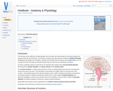
The hind brain is also called the rhombencephalon and is the brain …
The hind brain is also called the rhombencephalon and is the brain stem that provides the connection between the spinal cord and the rest of the brain. The hind brain contains many vital structures including the Medulla Oblongata, the Pons (the link between the cerebellum, forebrain and mid-brain) and the majority of the cranial nerves, III to XII. In general the brain stem governs essential functions that are carried out sub-consciously via reflexes.
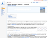
Hindgut fermenters are evolved to eat a herbivorous diet. Such a diet …
Hindgut fermenters are evolved to eat a herbivorous diet. Such a diet includes large quantities of insoluble plant carbohydrates, such as cellulose. Mammals cannot digest these insoluble carbohydrates as they lack the essential enzymes, such as cellulase. However it is important that they do digest these carbohydrates as there is insufficient quantity of soluble carbohydrates in plant material. Some microbes do have the enzymes to digest these insoluble carbohydrates and so hindgut fermenters hold a symbiotic relationship with these microbes. Hindgut fermenters have anatomical adaptations to allow for an expanded microbial population. The products of fermentation are volatile fatty acids. It is important to supply a source of fibre in their diet as it stimulates peristalsis in the gut and prevents a build up of gas.
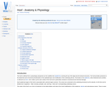
The hoof is defined from a physiologic perspective as the modified skin …
The hoof is defined from a physiologic perspective as the modified skin covering the tip of the digit and all enclosed structures. The hoof provides protection to the distal limb and is formed by keratinisation of the epithelial layer and modification of the underlying dermis. The keratin in the epidermis, when thickened and cornified, is referred to as horn. Horn makes up the outer surface if the hoof and is particularly resistant to mechanical and chemical damage.
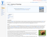
The keratin in the epidermis, when cornified and thickened, is referred to …
The keratin in the epidermis, when cornified and thickened, is referred to as horn. Horn is particulary resistant to mechanical and chemical damage. The dermis of horn gives the structures their 3-D structure and shape. Cattle, some sheep, goats and antelope posess horns and these are permanent organs. Breeds without horns are termed polled breeds. Deer posess antlers, which are temporary organs that develop during the rutting season and are then shed.
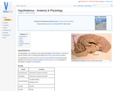
The hypothalamus is a small area in the ventral diencephalon of the …
The hypothalamus is a small area in the ventral diencephalon of the forebrain, in the floor of the third ventricle, and is a functional link between the nervous and endocrine systems.
The hypothalamus is inherently female. Testosterone 'defeminizes' the brain during embryogenesis and …
The hypothalamus is inherently female. Testosterone 'defeminizes' the brain during embryogenesis and eliminates the GnRH surge centre in males. The female foetus has no testes to produce testosterone, thus developes a hypothalamic GnRH surge centre.
The ileum is the terminal portion of the small intestine and continues …
The ileum is the terminal portion of the small intestine and continues from the jejunum. It opens into the caecum at the ileocaecal orifice. The intestinal epithelium is mainly absorptive, with much less digestion occurring compared to the duodenum and the jejunum.
The term 'implantation' is often used to describe the attachment of the …
The term 'implantation' is often used to describe the attachment of the placental membranes to the endometrium in most animals. True implantation is a phenomenon in rodents and humans in which the conceptus 'buries' itself in the uterine endometrium. The conceptus temporarily disappears beneath the surface. In most other species, the conceptus does not truly implant, but attaches to the endometrial surface and remains in the luminal compartment.
The integumentary system is an organ system that forms the protective covering …
The integumentary system is an organ system that forms the protective covering of an animal and comprises the skin (including glands and their products), haircoat or feathers, scales, nails, hooves and horns. The integumentary system has a variety of functions; in animals, it serves to waterproof, cushion and protect the deeper tissues, excrete waste, regulate temperature and is the location of sensory receptors for pain, pressure and temperature. Generally mammalian skin is covered with hair and is termed hirsute skin. Where hair is absent, it is termed glabrous skin.
The intermediate mesoderm exists as a strip of tissue between the lateral …
The intermediate mesoderm exists as a strip of tissue between the lateral plate mesoderm and somites. It gives rise to the urinary system and some parts of the reproductive system. Kidney development includes three forms: Pronephros, Mesonephros, and Metanephros. Mammals develop all three, and continue to use the metanephros in adult life. More primitive animals have only the first one or two.
Sexual differentiaton by default follows a pathways for development of female internal …
Sexual differentiaton by default follows a pathways for development of female internal and external genitalia, requiring no active intervention. Endocrine activity of the testes, production of Androgens by Leydig cells and Mullerian Inhibiting hormone (MIH) by Sertoli cells, is required for sexual differentiation to be diverted down the male genitalia development pathway.
The jejunum continues from the duodenum and leads into the ileum. It …
The jejunum continues from the duodenum and leads into the ileum. It is the longest part of the small intestine and is highly coiled. It has digestive and absorptive functions.
The kidneys are formed from the intermediate mesoderm. Firstly it begins to …
The kidneys are formed from the intermediate mesoderm. Firstly it begins to thicken longitudinally in its caudal domain. This is called the nephrogenic thickening. From this thickening three so called attempts at producing excretory origins arise. All the attempts have at the histological level a series of excretory tubules however only one of these attempts forms the kidney in adult life.
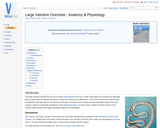
The large intestine extends from the ileum of the small intestine to …
The large intestine extends from the ileum of the small intestine to the anus. Water, electrolytes and nutrients are absorbed which concentrates the ingesta into faeces. Faeces are stored prior to defeacation. There is no secretion of enzymes and any digestion that takes place is carried out by microbes. All species have a large microbial population living in the large intestine, which is of particular importance to the hindgut fermenters. For this reason, hindgut fermenters have a more complex large intestine with highly specialised regions for fermentation.
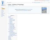
The larynx is situated below where the pharynx divides into the trachea …
The larynx is situated below where the pharynx divides into the trachea and the oesophagus. It is contained partly within the rami of the mandible and extends caudally into the neck. Vocal folds and vestibular folds are present in the larynx and due to this, it is more commonly known as the voice box.
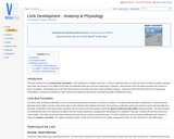
The limbs develop from the lateral plate mesoderm. Limb development is highly …
The limbs develop from the lateral plate mesoderm. Limb development is highly conserved; in all land vertebrates there are only four limbs and they are always opposite each other with respect to the midline of the body. All vertebrate limbs have the same patterning of; stylopod - proximal part of the limb which produces the humerus or femur; zeugopod - intermediate part of the limb which produces the radius and ulna or tibia and fibula; autopod - distal part of the limb that produces the carpals and metacarpals or tarsals and metatarsals. Other animals also follow this limb pattern including the greatly modified bird's wing.
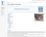
The liver (hepar) is an extremely important organ in the body of …
The liver (hepar) is an extremely important organ in the body of mammals and vertebrates as it provides functions essential for life. It is the largest internal organ and has numerous functions including production of bile and protein, fat and carbohydrate metabolism. During foetal development, the liver has an important haemopoetic function, producing red and white blood cells from tissue between the hepatic cells and vessel walls.
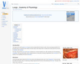
The lungs are the site for gaseous exchange, and are situated within …
The lungs are the site for gaseous exchange, and are situated within the thoracic cavity. They occupy approximately 5% of the body volume in mammals when relaxed, and their elastic nature allows them to expand and contract with the processes of inspiration and expiration.
No restrictions on your remixing, redistributing, or making derivative works. Give credit to the author, as required.
Your remixing, redistributing, or making derivatives works comes with some restrictions, including how it is shared.
Your redistributing comes with some restrictions. Do not remix or make derivative works.
Most restrictive license type. Prohibits most uses, sharing, and any changes.
Copyrighted materials, available under Fair Use and the TEACH Act for US-based educators, or other custom arrangements. Go to the resource provider to see their individual restrictions.